
By Assoc. Prof. Dr. Rania Hussien Al-Ashwal
Unlike other sectors, advancement in the medical field normally takes a while before reaching the consumer and reaching its actual applications in hospitals and clinics. This is not because there are fewer innovations made in the medical field, but rather the clinical trials that are generally required. After all, the implications of such innovations are for human lives. Much like data analytics, general stages of medical treatment start with diagnostics (finding out what went wrong and why) before prescription (providing countermeasures to treat the problem).
Accurate diagnostics would not be possible without technologies that enable insights, coupled with experiences with actual patients. This includes aortic dissection ᅳ a tear in the aorta, the large artery coming out of the heart ᅳ because if it is not properly diagnosed and intervention made promptly, it carries an attendant fatality.
Fortunately, real-life practice is one of many ways to gain experience, as it could also be gained through training with previous case studies including the study of medical records of patients as well as simulated cases. While the latter is often treated as “second-class” citizens, the field of simulations is scientifically valid. It is frequently used especially where the samples or the conditions are hard to obtain in real life. In Universiti Teknologi Malaysia (UTM), one such effort is the development of tissue-mimicking phantoms, specifically a model of imitation of the aorta of a person, to simulate both a healthy and diseased state.
The project lead, Associate Professor Dr. Al-Ashwal said, “We intended to develop a realistic training tool that enables health professionals to sharpen their diagnostic skills in a safe, controlled environment before they are faced with live cases.”
The researchers have made use of additive manufacturing, an advanced 3D printing technology to produce models of the aorta based on detailed body scanning, which can be used for practice in ultrasound diagnostics. Few materials were tested to determine which would best exhibit the echogenicity of human tissue under an ultrasound (one of the techniques used to detect aortic dissection).

“In our most recent research, we managed to create anatomically realistic aortic dissection simulators using polyvinyl alcohol (PVA) and silicon (Si) that effectively replicate the surface morphology and echogenicity of an actual aortic dissection.”
")
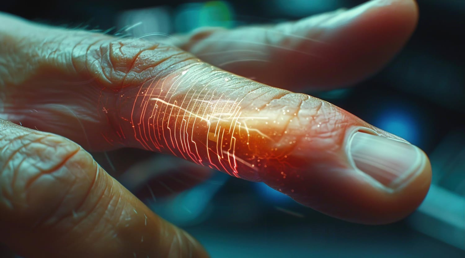
The same team also ventured into wound monitoring using the pioneering work of Nobel Laureate Charles Kuen Kao; fiber optics. Heavily used nowadays as communication tools, fiber optics has since found its way to various fields, primarily as sensors. As the light travels through fiber optics, the presence of different materials will affect how the lights behave and those behaviors can be detected and observations can be derived.
Applying fiber optic displacement sensors to decode the optical subtleties of human tissue, Associate Professor Dr. Al-Ashwal and her team applied them in working on an application to refine a refractive index that was an important measurement for analysis. “The refractive index,” Associate Professor Dr. Al-Ashwal explains, “is a window to the constitution of the tissue giving us a noninvasive means of gauging and tracking its condition, and that is of prime importance towards furthering patient care.”
One of the studies by Associate Professor Dr. Al-Ashwal revealed that the sensor (fiber optics) was able to distinguish between skin, muscle, and fat tissues, and to that extent, proving that there is a potential for this to be pushed into further investigation to determine the distinction between normal issues and abnormal tissues.

The medical field does not always need to reinvent itself. It would not be a surprise that inventions developed by other fields were found to be extremely useful in the medical field. As the world is moving at a break-neck speed, the medical field needs to continuously innovate to improve its effectiveness and efficiency. As the challenges of the world are getting more complicated to solve, engineers have a significant role to play, engaging in more transdisciplinary research and translating engineering applications into solutions.
Associate Professor Dr. Al-Ashwal’s works can be found in issue 24 of the Journal of Ultrasonography (doi: 10.15557/JoU.2024.0002) and issue 7 of the Journal of Optical Fiber Technology (doi: 10.1016/j.yofte.2024.103840).
Co-authors: Mutiah Rahmah, Thivya Nathini a/p Naderjan, Nur Syakirah Mohamad Safri
Edited by: Chew Teong Han
